
Powered By



A mammogram can reveal important clues about breast health, but sometimes the picture isn’t as clear as it could be. If you’ve been told you have dense breast tissue, you may wonder what that means for your mammogram and your breast cancer-screening plan.
This article explains what dense breast tissue is, why it can make mammograms harder to read, how it may affect breast cancer risk, and when other screening tests may be worth discussing with your doctor.
Dense breast tissue is a term used to describe how your breast tissue looks on a mammogram. It doesn’t mean your breasts feel firm or lumpy. You usually can’t tell whether you have dense breasts by touch or by breast size. Breast density is found only through breast imaging.
Breasts are composed of different types of tissue, including:
Dense breast tissue means there’s more glandular and fibrous tissue than fatty tissue.
Dense breasts are common. Almost half of women over 40 who get mammograms have dense breasts.
Breast density is usually described in four categories:
Almost entirely fatty — The breast has mostly fatty tissue.
Scattered areas of density — There’s mostly fatty tissue, with some dense areas.
Heterogeneously dense — There are many areas of dense tissue.
Extremely dense — Most of the breast tissue is dense.
The last two categories are considered “dense breasts.” About 4 in 10 women have heterogeneously dense breasts, and about 1 in 10 have extremely dense breasts.
On a mammogram, dense breast tissue appears white, while fatty breast tissue appears darker.
Dense tissue matters for two main reasons. First, it can make breast cancer harder to see. Second, dense tissue is linked with a higher chance of developing breast cancer.
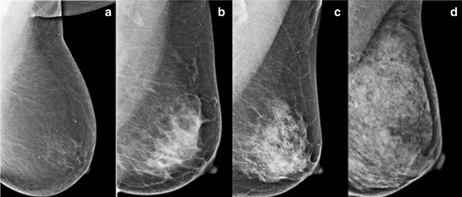
Dense breast tissue can mask breast cancer on a mammogram. Dense breast tissue appears white and so do some breast cancers and other abnormal changes. This overlap can make it harder for a radiologist (a doctor who reads imaging tests) to spot a tumor.
This is called “masking,” because the dense breast tissue overlaps, or hides, a concerning spot. A mammogram of more fatty breasts is often easier to read because fatty tissue appears dark and has better contrast with white breast changes.
Dense breasts lead to a reduction of 10 percent to 20 percent in the sensitivity of the mammogram. While about 80 percent of breast cancers can be detected by mammogram in women without dense breasts, the number decreases to below 60 percent for women with dense breasts, according to Mayo Clinic. The more dense the breast tissue, the lower the detection rate falls.
Dense breast tissue isn’t only a screening issue. It’s also an independent risk factor for breast cancer. That means people with dense breasts have a higher chance of developing breast cancer than people with mostly fatty breasts, even apart from how hard a mammogram is to read.
Researchers don’t know exactly why dense tissue raises breast cancer risk. It may be because dense breasts simply have more cells that could turn cancerous.
Breast density is one piece of your overall risk picture. Having dense breasts doesn’t automatically mean you’ll develop breast cancer. It means you may need a more personalized screening discussion with your doctor.
In the United States, mammography facilities must tell people whether their breast tissue is dense or not dense. This federal rule took effect Sept. 10, 2024.
Your report may say that dense breast tissue can make cancer harder to find and can raise breast cancer risk. It may also suggest talking with your doctor about breast density, your personal risk, and whether other imaging tests could help.
If you’re not sure what your report means, ask your doctor or the imaging center to explain your breast density category.
A mammogram is still an important screening tool for people with dense breasts. Even when dense tissue makes the image harder to read, mammograms can still find cancers and early warning signs. They can detect tiny calcium deposits, called calcifications, and other changes that may point to cancer.
A mammogram revealing dense breast tissue doesn’t mean the test “failed.” It means the test gave useful information, including breast density, but it may not tell the whole story for everyone.

Women with dense breasts may be more likely to discuss supplemental imaging after a mammogram than those with more fatty breasts. Supplemental screening may also be discussed based on breast density and overall breast cancer risk.
Some medical groups and specialists still suggest that extra screening can make sense for certain people, especially if you have dense breasts and a higher overall risk, such as:
The best choice depends on your risk level, what tests are available near you, and the possible downsides like false alarms.
Supplemental screening may include an ultrasound, MRI, or other types of imaging.
A breast ultrasound uses sound waves to make images of breast tissue. It may find some cancers that aren’t visible on a dense breast mammogram, which is why it’s one of the most commonly used supplemental screening tests for dense breasts.
One MyBCTeam member reported, “Mammo failed to find my cancer due to my dense tissue. Diagnostic ultrasound saved my life.”
One drawback is that ultrasound can lead to false positives. A false positive means the test finds something that looks concerning but turns out not to be cancer. This can lead to more imaging or a biopsy.
A breast MRI uses magnets and contrast dye to create detailed breast images. MRI can be especially useful for people with a higher-than-average breast cancer risk. The American Cancer Society recommends a yearly breast MRI plus mammogram for certain high-risk women, starting around age 30.
MRI can see through dense tissue better than mammography in some cases. However, it can also miss some cancers that mammography finds. That’s why it should be used in addition to, not instead of, a screening mammogram.
Some people with dense breasts may have 3D mammography, also called digital breast tomosynthesis. This test takes images from different angles, which may help doctors see through overlapping areas of tissue and may improve cancer detection, including in dense breasts.
3D mammography is becoming more common, but it isn’t available in all breast imaging centers. Also, it may cost more than a standard mammogram, so it may not be covered by insurance.
Some centers may offer contrast-enhanced mammography, molecular breast imaging, or abbreviated breast MRI. These tests may be options in certain situations, but availability varies by location.
Your doctor can help you decide whether one of these tests fits your risk level, health history, and local screening options.
 What Questions Should You Ask Your Doctor?
If your report mentions dense tissue, you can call your doctor’s office or the breast imaging center and ask the following questions:
It may also help to ask for a formal breast cancer risk assessment. This can include your age, breast density, family history, biopsy history, and other factors.
On MyBCTeam, people share their experiences with breast cancer, get advice, and find support from others who understand.
Have you been told you have dense breast tissue? Did it change your breast screening plan? Let others know in the comments below.
Get updates directly to your inbox.





 Continue with Facebook
Continue with Email
Continue with Facebook
Continue with Email
Continue with Facebook
Continue with Email
Continue with Facebook
Continue with Email
Become a member to get even more





 Join
Join



 Your Privacy Choices

This is a member-feature!
Sign up for free to view article comments.
It did in my case, and it didn't show up on an ultrasound either. This is probably the reason I was diagnosed at stage 4. Luckily I had calcifications that prompted a biopsy before it could spread… read more
We'd love to hear from you! Please share your name and email to post and read comments.
You'll also get the latest articles directly to your inbox.